Wherein it is proposed that spherules were fleshy masses of archaeobacteria growing in the ground like nodules on a root. These fleshy spheres were later replaced by hematite to become the spherules.
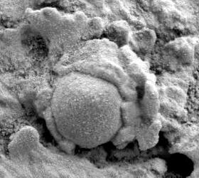
The spherules were found to be made of hematite, a form of iron oxide (Fe2O3). Hematite almost always forms in water. This analysis was confirmed by all three rover instruments, the mini-TES, Mossbauer, and Alpha Proton X-ray Spectrometers. The analysis was made in the "berry bowl" in the middle of March 2004, a shallow depression in the bedrock where dozens of spheres had collected together. While the surrounding bedrock does not show any sign of hematite, the spherules, gathered together in the "berry bowl" showed a very strong signal. While not pure hematite, the spherules are primarily formed from that mineral. The spherules are not blue, as they are sometimes depicted, but rather gray. The spherules tend to a normal maximum size, which suggests that they are biological and could be fossils, but they also have an even distribution across their mother rock which is bad for fossil theory.
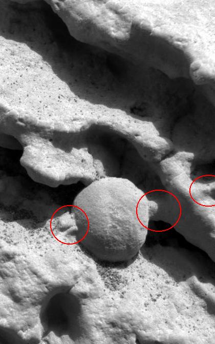
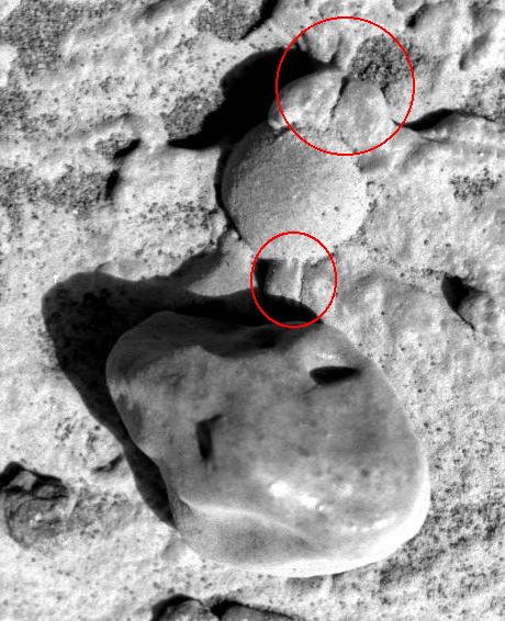
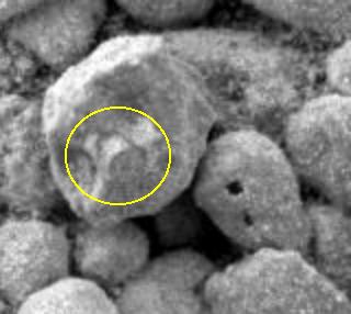
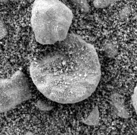
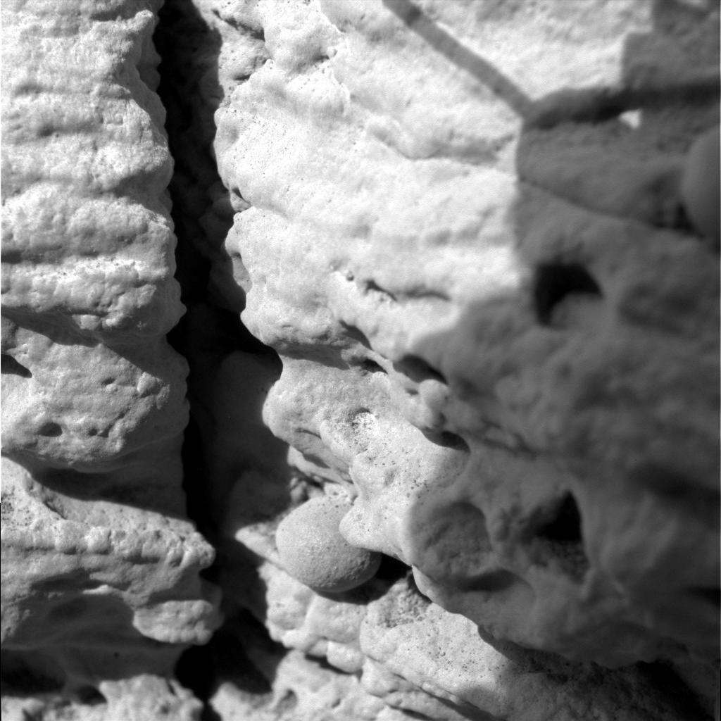
1./ Spherule in situ showing attached and penetrating axle or chord.
The chord extends 5 mm to the right and 1 mm to the left of the 4 mm diameter spherule.
A couple of "fingers" hold the chord to the rock.
If you squint it looks like the chord is possibly segmented like a crinoid's stem.
Notice the alternate sedimentary layers, one layer hard and cemented by
"primordial slime/algae/ooze/protoplasm",
and the next layer soft, eroded and sandy.
There is a curious hole directly below the spherule.
Opportunity :: Microscopic Imager :: Sol 015.
m/015/1M129515505EFF0312P2933M2M1
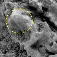
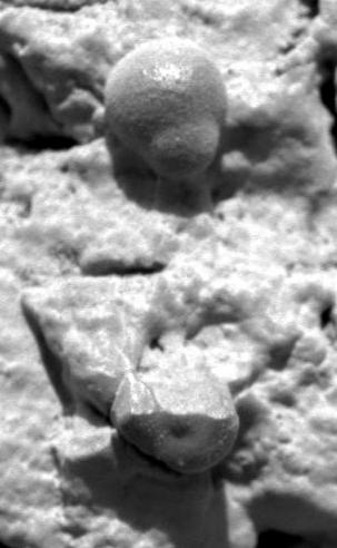
2./ Cross section of a spherule after grinding. Notice the stem.
m/034/1M131212783EFF0500P2959M2M1
The same spot before RAT abrasion - this spherule is the one in the upper right.
m/034/1M131201618EFF0500P2933M2M1
Opportunity :: Microscopic Imager :: Sol 034.
If these are fossils, which is possible but far from certain, then it is likely
these organisms evolved during the watery epoch of Mars hundreds of millions of
years ago.
Everything is ambiguous and nothing uncertain.
There is a shallow sulphur-rich sea.
The floor is covered by a microbial slime.
Everywhere tiny hematite spherules grow and these are connected by roots.
Or were the nodules of something else, some kind of suphur starch and
this was replaced later by hematite?
Are there other mineralized structures, our exo-cephalopods, exo-corals and worms?
This ancient eroded detrius is obscure and hazy and viewed through a glass darkly.
Wild speculation --
The spherules are made of hematite, a form of iron oxide (Fe2O3).
Both iron and carbon are dissolved in the ancient sea.
Some unknown type of martian photosynthesis takes two carbon dioxide molecules,
four dissolved iron atoms, and captures the needed carbon and
disposes of the iron as hematite spherules.
Is there hematite in my martian algae mats -- did they run off the same type
of photosynthesis?
End of wild speculation.
There is a brilliant page here showing terrestrial fossils that have been replaced in hematite.
Hematite Fossils -- http://www.natureshop-gallery.com/marsfossils/hematitefossils.htm
An equally brilliant page is here showing
inorganic hematite concretions, actually calcite, but coated with hematite.
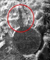
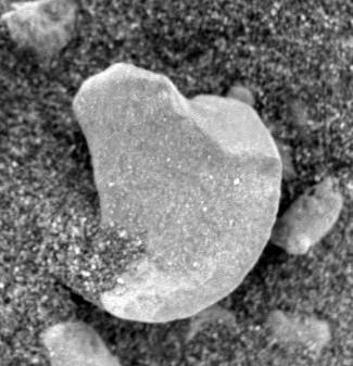
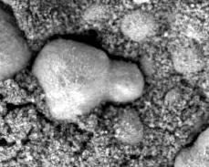
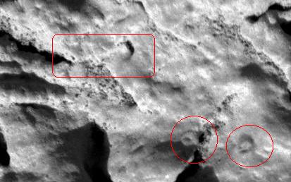
3./ Is this a fossilized root entering a spherule?
Are those other roots growing beside it?
It is rare for plant roots to fossilize.
Roots and stems are seen on fern fossils,
but then mostly as a black carbon outline and not solid as shown here.
Trees and branches fossilize.
If these Martian spherule stems were comparable to terrestrial sea weed stems,
that rarely fossilize, then it is odd that the Martian ones fossilized.
Notice both sides of the spherule are flatten but at an angle to each other.
There is a hole above the spherule.
Was that hole some kind of internal plumbing for the rock formation?
Opportunity :: Microscopic Imager :: Sol 040.
m/040/1M131734806EFF0544P2972M2M1
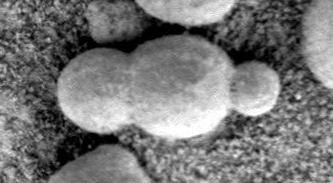
4./ A spherule with "funiculus" growing out two ends.
A "funiculus" is the stalk of a plant ovule or seed.
Spherule diameter = 4 mm.
Just ignore the cute little polished stone sitting on top.
Opportunity :: Microscopic Imager :: Sol 041.
m/041/1M131830596EFF0574P2952M2M1
|
5./ Two black-stained balls from excavated trench.
Notice the stem attachment point complete with central tap hole.
Opportunity :: Microscopic Imager :: Sol 025. m/025/1M130404506EFF0400P2953M2M1 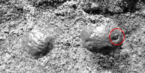
|
. |
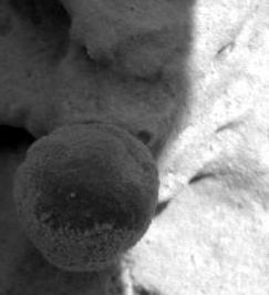
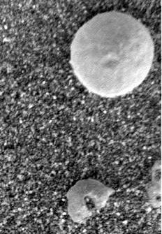
6./ Baby spherule with dimple.
Sol 10. From Jason. /1/m/010/1M129070954EFF0224P2933M2M1 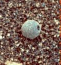
Second baby spherule with dimple.
Is there a trend here?
|
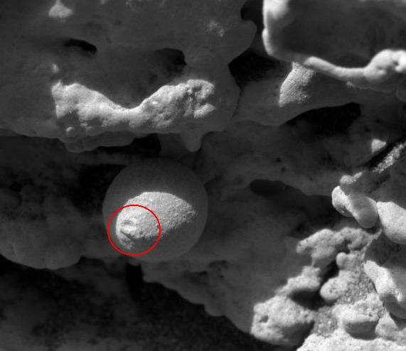
7./ A classic spherule with stem dimple. Spherule diameter = 4.2 mm.
You can see the core of the root going in.
This one is going to be in all the exobiology textbooks.
Opportunity :: Microscopic Imager :: Sol 039.
m/039/1M131649564EFF0544P2933M2M1
7.B/ Opportunity Sol 106.
An amazing shot of the spherule eroding away from a cylindrical inner core.
This cylindrical inner core is an extension from my hypothesized root and dimple.
This is a bit bizarre.
Sometimes you see the root dimples as craters because the root is softer and it erodes out.
From Hamdi Ucar of Turkey.
Enlarged double size.
The stone it is in is 2.5 mm in diameter. The cylindrical core is about .7 mm diameter.
Beside it is a detail of two oddly eroded rocks from the same picture.
/1/m/106/1M137593512EFF2208P2956M2M1
7.C/ Opportunity Sol 125.
From outside the Endurance crater.
The outer spherule has eroded away leaving the cylindrical inner core
complete with the inner-most tube.
/1/m/106/1M137593512EFF2208P2956M2M1
Three non-biological explanations for the dimples are proposed. Explanation #1 is the best and the other two are less satisfactory.
 Dimple explanation #1 - non-biological:
A feeder canal of mineralized fluid leads to each concretion.
The concretion grows in diameter and envelopes the feeder canal.
The spherule breaks away from the feeder canal and the dimple is
the root of feeder canal leading into the spherule.
The observations that support this theory and which the theory seeks to explain are
the plumbing holes in the mother rock around the spherule and vugs,
the dimples and the holes in the spherules,
the "golf tee" and other fossilized stalks,
the central cylindrical core found in some spherules, and
the roots and stalks and "golf tee" stalks, being in fact the mineralized canals,
leading to some in situ spherules.
Important point -- as shown later on in this article, spherules have an
hard outer crust. The interaction between the outer crust and the
mineralized canal may contribute to the remarkable shape of the dimple.
Dimple explanation #1 - non-biological:
A feeder canal of mineralized fluid leads to each concretion.
The concretion grows in diameter and envelopes the feeder canal.
The spherule breaks away from the feeder canal and the dimple is
the root of feeder canal leading into the spherule.
The observations that support this theory and which the theory seeks to explain are
the plumbing holes in the mother rock around the spherule and vugs,
the dimples and the holes in the spherules,
the "golf tee" and other fossilized stalks,
the central cylindrical core found in some spherules, and
the roots and stalks and "golf tee" stalks, being in fact the mineralized canals,
leading to some in situ spherules.
Important point -- as shown later on in this article, spherules have an
hard outer crust. The interaction between the outer crust and the
mineralized canal may contribute to the remarkable shape of the dimple.
Dimple explanation #2 - non-biological: Assume that the spherules are concretions and two of them grow closely together and the outside surfaces touch. There is a weakness at the point of contact and when the two spherules separate a dimple is formed on each one. Further, impure hematite is a poor conductor, but it is still a conductor. With two hematite spherules leaning against each other there be some electrochemical interaction at the point of contact? From Telford et al. (1976), "Hematite and sphalerite, in pure form, are practically insulators, but when combined with impurities may have resistivities as low as 0.1 ohm-m." The stalks or roots shown in the pictures above would be a mineralized feeder canal for the spherules, or maybe a cross section of the sedimentary layer the spherules like to form on. Against this explanation we can say that the dimple seems to form on a spherule pole and you never see two or more dimples, derived from random contacts, on the same hemisphere.
Dimple explanation #3 - non-biological: The spheres are formed as a type of integral spherical crystal. The placement of hematite molecules around the surface of a sphere is not regular. A singularity develops around the poles. It is at the poles where the crystal is the weakest, that the dimple forms when the erosion starts. This explanation is not really convincing as the uneroded spherules have bumps and grooves and textured surfaces and impure interiors.
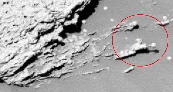
8./ Two spectacular images from Panoramic camera sol 37 and 16.
Spherules eroded with roots showing. Enlarged two times.
I first saw these pictures on an extinct site fellow Canadian Paul Anderson made about Mars.
Opportunity :: Panoramic camera :: Sol 037.
These would have made a nice cover picture for National Geographic so it is surprising
that NASA didn't image them more closely.
p/037/1P131476754EFF0518P2392L7M1 and sol 16
p/016/1P129617432EFF0322P2260L7M1
|
From the panoramic camera, sol 85.
p/085/1P135731395ESF1400P2532R1M1
Compare with the
simple erosion protection streaks of /p/085/ and the
golf tees of /p/086.
Look also at this image of the
mineralized "golf tee" channel from sol 086 - m/086/1M135817872EFF1407P2936M2M1
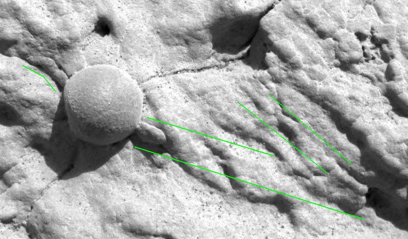
What are the "golf tee" stalks or peduncles? Are they: 1./ the remains of biological roots, or 2./ some kind of rocky plumbing channel that feeds a mineralized fluid to the concretion and is harder than the surrounding matrix and erodes out slower, or 3./ an erosion phenomenon with the rock matrix protected from the wind by the harder spherules? Following the "potato on root" biological fossil theory, we would want the stalks on both sides of the spherule. But they only appear on the back. The golf tee stalks are most likely a combination of #2 and #3. The direction the stalks point in is controlled half by the rock surface and half by weathering from prevailing winds. The stalks seem to be co-planar with the sediments layer. Did the concretion enlarge to grow around the mineralized feeder channel "golf tee" stalks and break off, forming the dimples including the root into the dimples? Hmmm...
Compare the spherules on stalks with these concretions,
probably non-biologically produced, shown in the two following articles. |
|
|
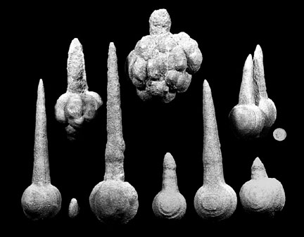
The Spherules and the Horodyskia:
There is a little known terrestrial biological fossil parallel to the spherules.
This is the horodyskia, the 1.5 billion year old 'string of beads' fossils
found in Glacier National Park, Montana.
There is also a Western Australia species whose beads are not so large as some from Montana.
They are presented as the oldest known tissue-grade colonial eucaryote.
Their nature is uncertain but they could be
exceptionally large bacterial communities, or megascopic seaweeds,
or even animal trace fossils.
The horodyskia fossils preserve both the beads and traces of the string connecting the beads.
For more about the horodyskia, see New occurrences of �strings of beads� in the Bangemall Supergroup: a potential biostratigraphic marker horizon by K. Grey1, I. R. Williams1, D. McB. Martin1, M. A. Fedonkin2, J. G. Gehling3, B. N. Runnegar4, and E. L. Yochelson. "The strings of beads consist of impressions of 3 to 30 beads, in strings from 1 to 7 cm long Bead diameter ranges from 0.5 to 4 mm. Impressions are up to 1 mm deep and typically between 1 and 5 mm apart." Also see The dawn of multicellular life: evidence from the Bangemall Supergroup by D.McB.Martin. |
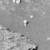
 Terrestrial Horodyskia. Image from Monash University -- School of Geosciences. Used without permission.
|
|
|
Wild speculation: One and a half billion years ago a giant meteor smashes into the Earth which is at this time mostly inhabited by Horodyskia. The tremendous explosion blasts pieces of rock, some with Horodyskian bacteria, into space where the rocks' trajectory takes them to Mars. These terrestrial meteorites crash down onto Mars, break apart and release their bacteria that has miraculously survived the trip through space. The wet environment of Mars is suitable for the bacteria and soon the planet is host to a thriving colony of them. These evolve into the fleshy spheres that will later be replaced by hematite and become our spherules. The spherules are direct descendents of the Earthly Horodyskia. Fast-forward now one and a half billion years. An amateur naturalist discovers the fossil nature of the spherules and has an epiphanic and vast vision of life arising independently throughout far-flung galaxies. Suddenly, on the eve of his 50th birthday, he becomes aware of the Horodyskian connection. He is suddenly plunged into despair as he realizes that one cannot philosophically project life onto other star systems, where the only known second biology case happens to be an accidental contamination from the unique case of Earth's life. If the objects described on this page are proved to be biological, then it is very possible that Mars's life was a result of contamination from Earth. It cannot be that easy for life to develop.
|
||
|
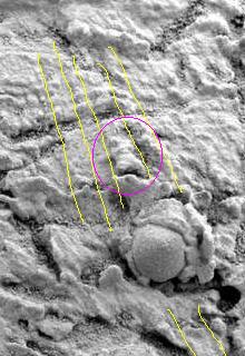
Did the Spherules Grow Originally in Lines?
Did our hypothesized spherical clumps of Martian archeobacteria
grow originally in lines and connected by threads
in situ in their mother rock like the Horodyskia?
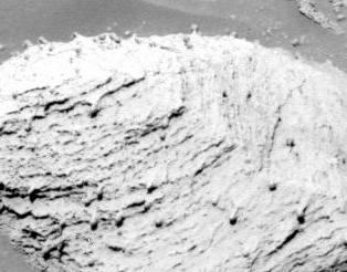
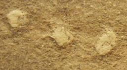
Certainly, after being eroded out, the spherules tend to be pushed, by some process operating over milleniums, into connected rows with one spherule leaning directly against the nest. This is shown in this http://www.lyle.org/~markoff sol 94 colour image. That same image shows spherules cemented into the ground into these same tight rows. Here we are looking down along the top surface of a single sedimentary plane. It looks like the first spherule gets anchored somehow, and over millenia, others are pushed by gusts of winds into the lee behind the anchored one. For the spherules growing in rows, each one in sequence is resting against the one in front and behind. This is true for the free ones, as well as the ones cemented into the ground. Marvi writes: "I saw some image, here on earth, of small rocks and cobbles in a cold desert, undisturbed for eons and aligned in lines and curves. I wish I could find it again. It seems that these alignments are a very common and well understood."
|
Are the spherules growing in rows like the horodyskia? Some of them look cemented in. From http://www.lyle.org/~markoff sol 94 colour image. |
Images of the Spherules in Situ:
The following images show the spherules eroding from their mother rock:
|
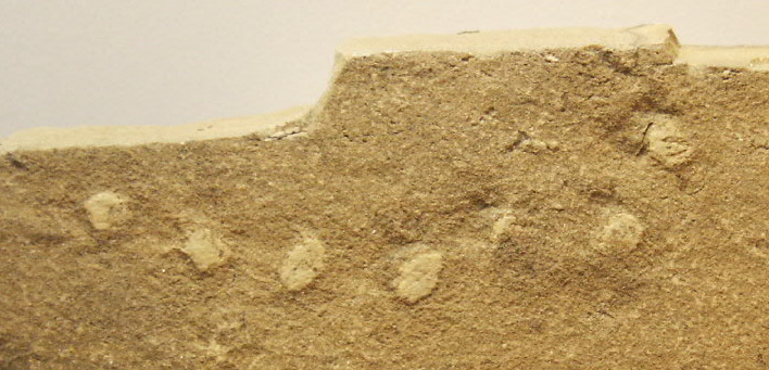
/1/p/118/1P138654324EFF2809P2298L2M1 In this image our spherules are presented nicely growing in lines on a single strata layer, each spherule separated by half an inch or so of free space just like with the Horodyskia. On the larger NASA image, is that an ancient rain water wash channel between the rocks? Could the wind do that over time? Opp sol 118 Pancam. |
|
| If the spherules grew along a mineralized feeder pipe, it would make sense that they would grow in rows. Maybe they grow solitary, and wave action pushes them into lines. Besides, nothing depends on them growing in rows. Maybe water currents breaks the spherules and their stems apart and they grow solitary after that. Their lines go in all directions and they are are not really heaped as wave action might do. Many of the newly exposed ones with the pristine surfaces do not show dimples that might come from leaning against a second spherule or from a feeder channel. | ||
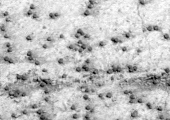
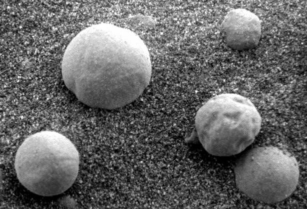
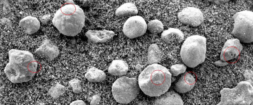
9./ Some newsgroup postings suggest spherules come in only one size. This is false.
Five balls showing their irregular sizes. The largest has "tennis ball" markings.
The one in the lower right is marked as well.
The largest is 5 mm in diameter. The smallest is 2 mm in diameter.
Opportunity :: Microscopic Imager :: Sol 014.
|
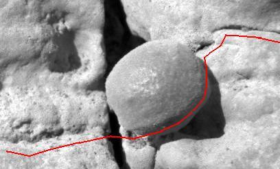
10./ Be cautious with marking and grooves on the spherules.
It may be that some grooves on the spherules represent something in the underlying rock.
For example, the ridge on this spherule follows the weak layer
in the rock that surrounded it as it was growing. From sol 039 /1/m/039/1M131656918EFF0544P2972M2M1. Having said that, I encourage readers to click here to view the very detailed study of the marking on the spherules by Albert de Hoogh of the Hague in the Netherlands. |
. |
|
10./ Another one with a line on it ressembling the seam of a tennis ball.
Opportunity :: Microscopic Imager :: Sol 039.
m/039/1M131650751EFF0544P2931M2M1 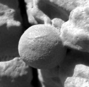
|
. |
A nice shot showing surface texture before weathering -- 3 mm diameter.
After they lie on the ground for a while they lose the fine details, their dimples and
the polish of their patina. Then they start to more like regular concretions.
Opportunity :: Microscopic Imager :: Sol 034.
m/034/1M131201618EFF0500P2933M2M1 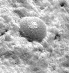
|
|
11./ A spherule in cross section with layering.
Usually spherules do not show any internal structure or layering, so this first one,
with a distinctive outer, a middle and an inner core is a bit of a novelty.
It may not be a spherule. It actually looks more cylindrical than spherical.
3 mm in diameter.
Opportunity :: Microscopic Imager :: Sol 028. m/028/1M130672724EFF0454P2933M2M1 There is another triangular "exo-cephalopod", maybe squashed a bit flat, behind it. Regarding the curious spherule above, did two spherules seed close together and grow into one another. |
. |
|
|
Cracked spherule showing possible outer layer. A big one -- almost 7 mm in diameter.
This picture is more typical of their appearance when broken and weathered.
Opportunity :: Microscopic Imager :: Sol 023. |
. |
|
|
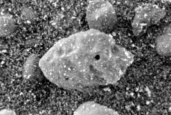
12./ What is this?
One of many odd-looking eroded pieces from the ground outside of the Eagle Crater.
This piece is 6 mm long which is very long for a spherule
and it looks like it might have been longer.
It seems that the outer layer was hard and the inside soft.
Maybe our proposed biological spherules have a crusty exterior and a soft interior.
Alternatively, concretions too, can have a hard crust and soft interior.
Have a look at the very interesting original picture.
the ground outside the Eagle Crater.
Opportunity :: Microscopic Imager :: Sol 056.
m/056/1M133157669EFF06HEP2956M2M1
|
. |
|
|
A second broken spherule.
Look carefully to see the harder outer layer. 4 mm in diameter.
Opportunity :: Microscopic Imager :: Sol 049.
m/049/1M132536343EFF0600P2956M2M1 |
. |
|
|
A crusty spherule from sol 86.
This one is held up above the surface and you can see the
mineralized "golf tee" channel leading to it as the softer rock has eroded away.
Sol 086.
m/086/1M135817872EFF1407P2936M2M1 |
. |
|
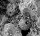
13./ Interwelded spherules. They are starting to look here like concretions.
Alternative wild speculation -- they are budding.
Opportunity :: Microscopic Imager :: Sol 052 and 46.
m/052/1M132808239EFF06A8P2956M2M1
and
m/046/1M132266828EFF05AMP2937M2M1
|
14./ Spherule root stem cross-section -- speculative. A lovely specimen only a millimeter in diameter.
Look at the menorah-like impression left at the bottom. Very nice sub-millimeter resolution. Opportunity :: Microscopic Imager :: Sol 2004-03-08. 2004-03-08/1M131655864EFF0544P2972M2M1 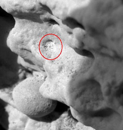
|
. |
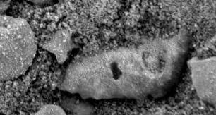
15./ Spherule root stem stub.
The circled object, 1 mm or so in diameter, the proposed stub,
looks like it may have had an internal tubular structure.
Beside it to the left is a stub that perhaps held a spherule once. Opportunity :: Microscopic Imager :: Sol 039. m/039/1M131652818EFF0544P2933M2M1 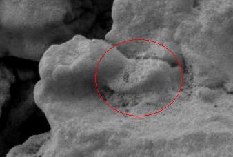
As with most of the pictures here, these two are somewhere in the "maybe" to "doubtful" continuum. |
16./ Are these spherule dimple stalks? Yes, no, maybe, definitely not. These are about 1 mm in diameter, comparable to the dimple diameters.
 |
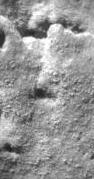
On the left: Opportunity sol 30.
This same area abraded gave us the Rover rotinis.
On the right:
Opportunity sol 105.
There is an eroded spherule at the bottom of this image.
I am likely stretching things here. We should put these with the doubtful images. |
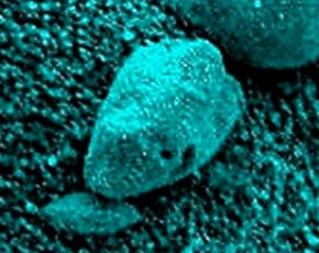
17./ An interesting and suggestive shot showing eroded and degraded spherules.
In a few places you can see how the stalk dimple has eroded differently
from the body of the spherule. The NASA image is a bit clearer, as usual.
The degraded spherule farthest to the right was pointed out to me, with careful analysis,
by Pedro Rosa of CIT KSU Kazan, Russia who provide the blue image below.
Opportunity :: Microscopic Imager :: Sol 049.
m/049/1M132536403EFF0600P2956M2M1
17B./ Another spherule with a tap hole -- 6 mm long.
Is this a weakness in the material where the stem has entered the spherule?
Some pebbles at your local beach have holes in them too, but
then again, at the local beach you have water freezing and thawing and
there are pebbles with organic shell pieces in the rock matrix.
Opportunity :: Microscopic Imager :: Sol 068.
m/068/1M134219950EFF08AQP2956M2M1
18./ An early dimpled spherule from February 5 2004. 4.5 mm diameter.
How many people looked at this and said, "I wonder if that is a fossil"?
These stem attachments to spherules observations have been made by many people
all over the Internet. I am not pretending I am the first person to observe these.
In all the newsgroups and bulletin boards people were making biological speculations
about them.
Opportunity :: Microscopic Imager :: Sol 034.
m/010/1M129070954EFF0224P2933M2M1
19./ A maddeningly out of focus shot of a dimple.
This could have been the "smoking gun" of spherule dimples.
There seems to be five radiating lines on the bedrock behind the spherule with the spherule at their centre.
Opportunity :: Microscopic Imager :: Sol 050.
m/050/1M132624683EFF0602P2956M2M1
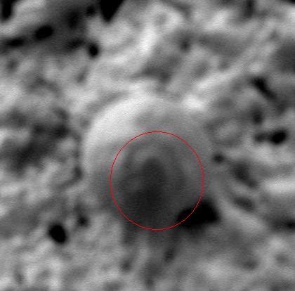
20./ Two views of the same spherule in situ from Opp sol 125 outside
the Endurance crater.
The feature circled in purple could be from the same sort of hardened feeder pipe
that causes the "golf tees" stalks. Traces leading to the spherule are shown in yellow.
Was there some kind of soft body growing around the spherules and cloaking it?
There were no indication of these cloaks on the spherules at the Eagle crater.
The nature of spherules suggested by this picture is completely different
from the nature of spherules suggested by the other pictures we have looked at.
/1/m/125/1M139280589EFF2829P2976M2M1
 |
 |
Opportunity :: Panoramic Camera :: Sol 029 -- viewing the fossiliferous rock.
Opportunity :: Navigation Camera :: Sol 026 -- The exposure of the fossiliferous rock.
Panoramic view of the "El Capitan" site where the fossiliferous rock is exposed.
A nice selection of panoramic camera images from sol 27.