The Sol 34 object
|
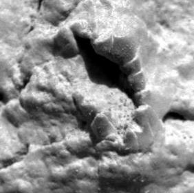
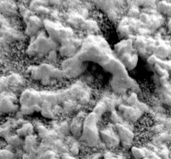
 The Sol 34 object -- which is more likely to be petrified mud than is a fossil.
This object is 6 mm high and 3 mm across.
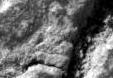
Compare the original rock surface
m/034/1M131201699EFF0500P2933M2M1
with the image from the same location only abraded
m/034/1M131212854EFF0500P2959M2M1.
On the abraded image a long dark arc of hard material
starts where the "fossil" object emerges on the outside of the rock,
and continues down to the bottom where there is curiously, a
chambered and/or segmented structure.
This structure is shown at the right and is 1.5 mm wide and 2 mm long.
There has been much speculation about this image, most of it with only entertainment value.
I would hesitate to call it a fossil.
If there were more pieces, if there were another piece of it bifurcating,
if this piece was longer, if the abraded image showed a fossil image, then maybe
otherwise no. Believe me - it is not a crinoid.
The Sol 34 object -- which is more likely to be petrified mud than is a fossil.
This object is 6 mm high and 3 mm across.
Compare the original rock surface
m/034/1M131201699EFF0500P2933M2M1
with the image from the same location only abraded
m/034/1M131212854EFF0500P2959M2M1.
On the abraded image a long dark arc of hard material
starts where the "fossil" object emerges on the outside of the rock,
and continues down to the bottom where there is curiously, a
chambered and/or segmented structure.
This structure is shown at the right and is 1.5 mm wide and 2 mm long.
There has been much speculation about this image, most of it with only entertainment value.
I would hesitate to call it a fossil.
If there were more pieces, if there were another piece of it bifurcating,
if this piece was longer, if the abraded image showed a fossil image, then maybe
otherwise no. Believe me - it is not a crinoid.
Opportunity :: Microscopic Imager :: Sol 034.
|
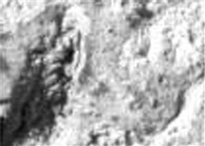
The Martian Rotini
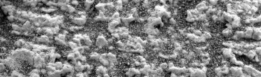
1./ Here is the famous rover rotini that was visible after a rock surface
was abraded by the RAT tool.
The rotini is very tiny, only 1.3 mm long and 0.1 mm wide --
not much more than specks of dust strung together.
There is another banded object just below and to the left --
it is maybe cross-section through one of the vug minerals.
The top two segments of our rotini, and the bottom one are not particularly
well preserved.
Its just those middle three segments, especially the middle one, that
gives it the impression of being a segmented organism.
On the other hand, segmentation is a feature of early life because
it is an easy way to reuse genetic material.
Compare the effect of focus and lighting of two images taken three sols apart - sols 030 and 33.
Sol 30 - /1/m/030/1M130859833EFF0454P2959M2M1
Sol 33 - /1/m/033/1M131117213EFF0454P2953M2M1
Here is the original unabraded surface for comparison -- sol 030.
The supposed spherule dimple stalks are in the upper left hand corner.
m/030/1M130846496EFF0454P2933M2M1.
|
Rotinis as abrasion artifacts:
Here is a similar-looking rotini-thingy created from the dust of grinding.
The original rotini looks much more solid and robust than the second rotini-thingy does.
It may be that the original rotini was an abrasion artifact but just
looking at the two, it would be a stretch to compare them
and say the original rotini was created by the grinding.
Even with harder rock and a deeper crack to fall in. Opportunity sol 108.
/1/m/108/1M137782381EFF2222P2956M2M1.
The abrasion dust rotini-thingy is about 2 inches over and 2 inches down from the top left corner of this image.
|
|
|
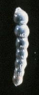
If these objects were fossils, it is possible that they were some sort of
martian marine foraminfera, single-celled protists with shells.
A vaguely similar looking terrestrial foraminferum - 2 mm long or so,
for what it is worth, is shown on the right.
A page showing other foraminferum is available at
Foraminifera from the Journal of Foraminiferal Research.
It might be also something like a
sponge spicule or gemmule or some other
tiny mineral piece, not the whole, of a marine animal.
Our fossil would not be a tiny 1.3 mm soft-bodied worm, sitting in a burrow no less,
for example,
as soft-bodied creatures are usually not fossilized under these conditions.
If the camera had caught a colony with dozens of these creatures, it would make
it easier to pronounce them as fossils -- with just one, the case becomes harder to make.
With foraminfera, we would expect to see dozens of them, just not one.
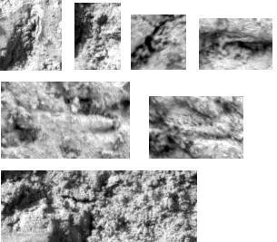
However as you study the image and the images of the hallucinigenia shown later,
you do see some similarly size structured objects,
but most of these are likely artifacts of high magnification.
Throughout different abraded areas several biological-looking
objects will appear if one stares at them long enough.
Some of these are shown in the next picture.
I encourage browsers interested in finding out more about rotinis
to study the original picture and better appreciate the play of
the magnified mineral and dust on the surface.
Here is a link to a rotini study image by Greg and Teresa Castleberry
of Bellevue Washington:
http://www.gtse.net/FossilMarkUp.jpg
Here is a
rotini study image
by Marvi.
|
|
Here is a second rotini candidate from sol 39. 1 mm long.
Rotini is a plural word, isn't it.
m/039/1M131655288EFF0544P2971M2M1
Vugs
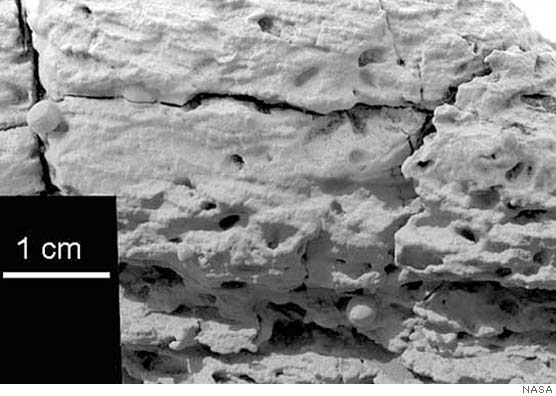
From the NASA press release of March 2, 2004.
"Pictures from the rover's panoramic camera and microscopic imager reveal the target rock, dubbed
"El Capitan," is thoroughly pocked with indentations about a centimeter (0.4 inch) long and one-fourth
or less that wide, with apparently random orientations. This distinctive texture is familiar to
geologists as the sites where crystals of salt minerals form within rocks that sit in briny water.
When the crystals later disappear, either by erosion or by dissolving in less-salty water,
the voids left behind are called vugs, and in this case they conform to the geometry of
possible former evaporite minerals."
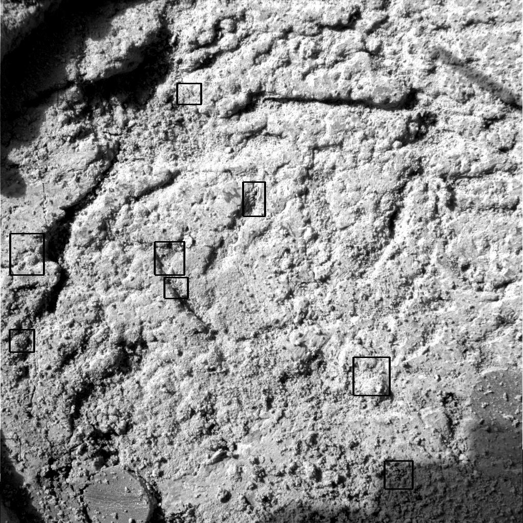
1./ An example of the famous vugs that the Opportunity lander found.
The "Y" and "V" shaped cavities represent negative space left over when
some hard structures, likely gypsum, were surrounded by clay and then dissolved out.
The question is whether at least some of the hard minerals later removed
were parts of a martian animal or plant.
Opportunity :: Microscopic Imager :: Sol 029.
m/029/1M130761192EFF0454P2933M2M1

It must have been a remarkable site for crystalizations what with both the
salt mineral vug crystals and the spherules slowly precipating out from the water.
Maybe it was a deep sea and as it slowly evaporated, the mineral concentration
became slowly saturated and the spherules and vug minerals precipated out.
The author, by the way, tends to agree with the orthodox scientific view that these hollows were
originally filled with some mineral, maybe gypsum, that was placed there non-biologically.
Click here for a great microscopic
vug panoramic collage.
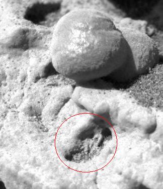
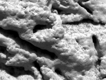 2./ Exo-cephalopod or a cone-shaped structure resembling a cephalopod.
This one is genuinely problematic and really looks like a fossil with
chambers no less. If "those were vugs that were its mouth", then possibly you
can say the yug cavity goes backwards into the rock while the shell goes
across flat against the rock. There are similar little triangular structures all
over the place although perhaps less dramatic as this one.
The structure is 10 mm long.
A serious problem with the fossil idea is that a cephalopod is a fairly advanced organism.
Terrestrial ones only developed in the Silurian, maybe 350 million years ago.
A martian exo-cephalopod implies martian life evolved to an advanced state and
that Mars remained a living planet up to a few hundred million years ago.
That would be an incredible parallel to make between martian and terrestrial life.
Organisms only evolve a hard shell when somebody with teeth is trying to eat them.
It is more credible to say this object is just an illusion caused by
magnification of the rock matrix shaped by surrounding vugs.
Opportunity :: Microscopic Imager :: Sol 029.
2./ Exo-cephalopod or a cone-shaped structure resembling a cephalopod.
This one is genuinely problematic and really looks like a fossil with
chambers no less. If "those were vugs that were its mouth", then possibly you
can say the yug cavity goes backwards into the rock while the shell goes
across flat against the rock. There are similar little triangular structures all
over the place although perhaps less dramatic as this one.
The structure is 10 mm long.
A serious problem with the fossil idea is that a cephalopod is a fairly advanced organism.
Terrestrial ones only developed in the Silurian, maybe 350 million years ago.
A martian exo-cephalopod implies martian life evolved to an advanced state and
that Mars remained a living planet up to a few hundred million years ago.
That would be an incredible parallel to make between martian and terrestrial life.
Organisms only evolve a hard shell when somebody with teeth is trying to eat them.
It is more credible to say this object is just an illusion caused by
magnification of the rock matrix shaped by surrounding vugs.
Opportunity :: Microscopic Imager :: Sol 029.
m/029/1M130761192EFF0454P2933M2M1
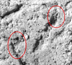
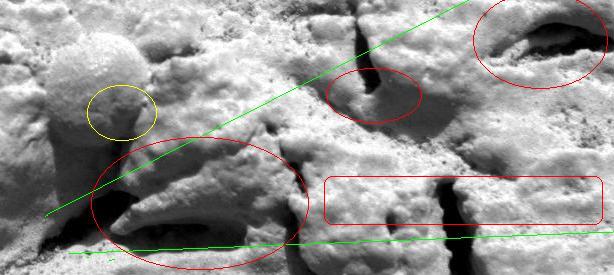
3./ Exo-cephalopod #2 or a cone-shaped structure resembling a cephalopod.
Green lines outline the proposed continuous organism. Red circles point out where
the organism wall was.
The structure is 15 mm long.
Or is this another example of a cone being formed between surrounding vugs.
In yellow, at no extra charge, a dimpled spherule with clefts.
Opportunity :: Microscopic Imager :: Sol 029.
m/029/1M130761497EFF0454P2953M2M1
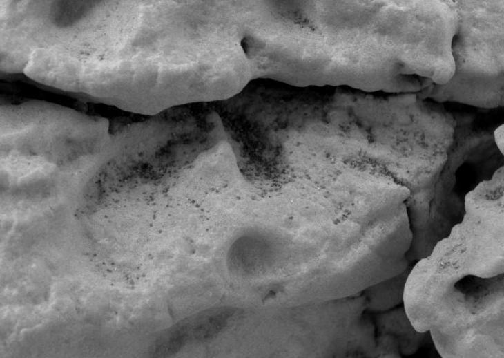
4./ Pseudotrilobite or eroded vug or Martian clam impression. 18 mm wide.
Why does it have a spine?
Are the holes above and below it related to it somehow?
The surface seems pitted? It is most likely an exposed vug --
the external form left when a crystal or mineral dissolves away.
Here is a
context picture
of this interesting specimen.
Note it is fives times larger than any of the vugs around it.
Opportunity :: Microscopic Imager :: Sol 039.
m/039/1M131651988EFF0544P2933M2M1
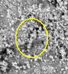
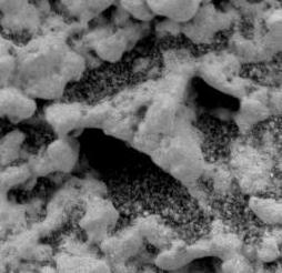
5./ Cone-shaped structure with distinct wall and cavity.
The outside of the elipse is about 5 mm long.
Once you start enlarging random crystals this much your eyes start playing tricks on you.
Look carefully. This is just a "vug" with crystals around the mouth.
Opportunity :: Microscopic Imager :: Sol 029.
m/029/1M130762907EFF0454P2953M2M1
|
. |
6./ Suggestive shape with distinct wall and cavity. 7 mm diameter.
Opportunity :: Microscopic Imager :: Sol 029.
|
7./ Elongated structure with wall??.
I would approach this one with caution.
This is the rock exposure that NASA said had water cross-bedding layers on it.
Actually, they were pointing exactly at this spot.
Still, I think it looks more than a bit like a cephalopod.
Opportunity :: Microscopic Imager :: Sol 028. 30 mm long.
m/028/1M130671865EFF0454P2953M2M1